Deep Learning Based Lung Image Segmentation Using XR-U-Net
Date:

In this talk, we explore the implementation of our research on “Deep Learning Based Lung Image Segmentation Using XR-U-Net”. This work focuses on enhancing the accuracy of lung image segmentation without preprocessing of images using a novel deep-learning architecture.
Introduction
Lung image segmentation is a crucial process in medical imaging. It involves isolating lung regions in X-rays or CT scans, aiding in diagnosing and treating lung diseases. Accurate segmentation allows medical practitioners to better visualize the lung structure and identify abnormalities.
Chest X-rays are one of the most accessible and cost-effective medical imaging methods globally, accounting for over one-third of all medical imaging. They are essential for diagnosing conditions like tuberculosis, pneumonia, and lung nodules. The U-Net architecture, with its skip connections, excels in medical image segmentation, achieving precise results.
Our Contributions
In our work, we:
- Developed XR-U-Net, an extended U-Net architecture with a five-layer encoder-decoder structure.
- Removed preprocessing steps to simplify the segmentation workflow.
- Conducted detailed visual evaluations of segmented images to validate performance.
Dataset
Our study utilized the Montgomery County chest X-ray dataset:
- 138 images in total, including:
- 80 normal cases.
- 58 cases with tuberculosis manifestations.
- The dataset was split into:
- 112 images for training,
- 13 for validation, and
- 13 for testing.
This dataset provides a foundational base for evaluating our model’s effectiveness.
System Architecture
The XR-U-Net system architecture enhances the traditional U-Net with a quintuple-layer encoder-decoder structure. This innovation allows for deeper feature extraction and improved segmentation without requiring preprocessing steps.
Metrics and Training
To evaluate performance:
- IoU (Intersection over Union) measures the overlap between the predicted and actual masks.
- Dice Coefficient quantifies the similarity between predicted and ground-truth masks.
The model training was carried out over 100 epochs using the Adam optimizer with fine-tuned parameters, achieving strong convergence as seen in the training curves.
Evaluation
The XR-U-Net achieved impressive results:
- Segmentation Accuracy: 95.7%
- IoU: 0.83
- Dice Score: 91%
Visual results showcase accurate lung boundary predictions that align closely with ground truth, as demonstrated in the comparison images.
Comparative Study
When compared with existing methods:
- XR-U-Net achieves a balance of accuracy and computational efficiency.
- While U-Net++ and Deep Residual U-Net achieved higher accuracy, their models were more complex and resource-intensive.
Limitations
Despite promising results, our model has some limitations:
- Limited dataset diversity restricts generalizability.
- Focused only on two-dimensional segmentation.
- Minor discrepancies in segmenting intricate lung structures.
- Lack of integration with advanced diagnostic frameworks.
Future Work
Future directions for this research include:
- Expanding datasets to include diverse cases.
- Exploring three-dimensional segmentation.
- Integrating the XR-U-Net model with diagnostic frameworks to identify conditions like cardiomegaly.
Conclusion
The XR-U-Net model demonstrates strong potential in advancing lung segmentation accuracy, achieving a Dice score of 91% and an IoU of 0.83%. Its simplicity and efficiency make it suitable for clinical applications, with the promise of further improvements in future work.
Acknowledgment
We extend our gratitude to the Department of Information and Communication Engineering, Pabna University of Science and Technology, for their support and funding of this research.
Feel free to contribute, raise issues, or suggest improvements!
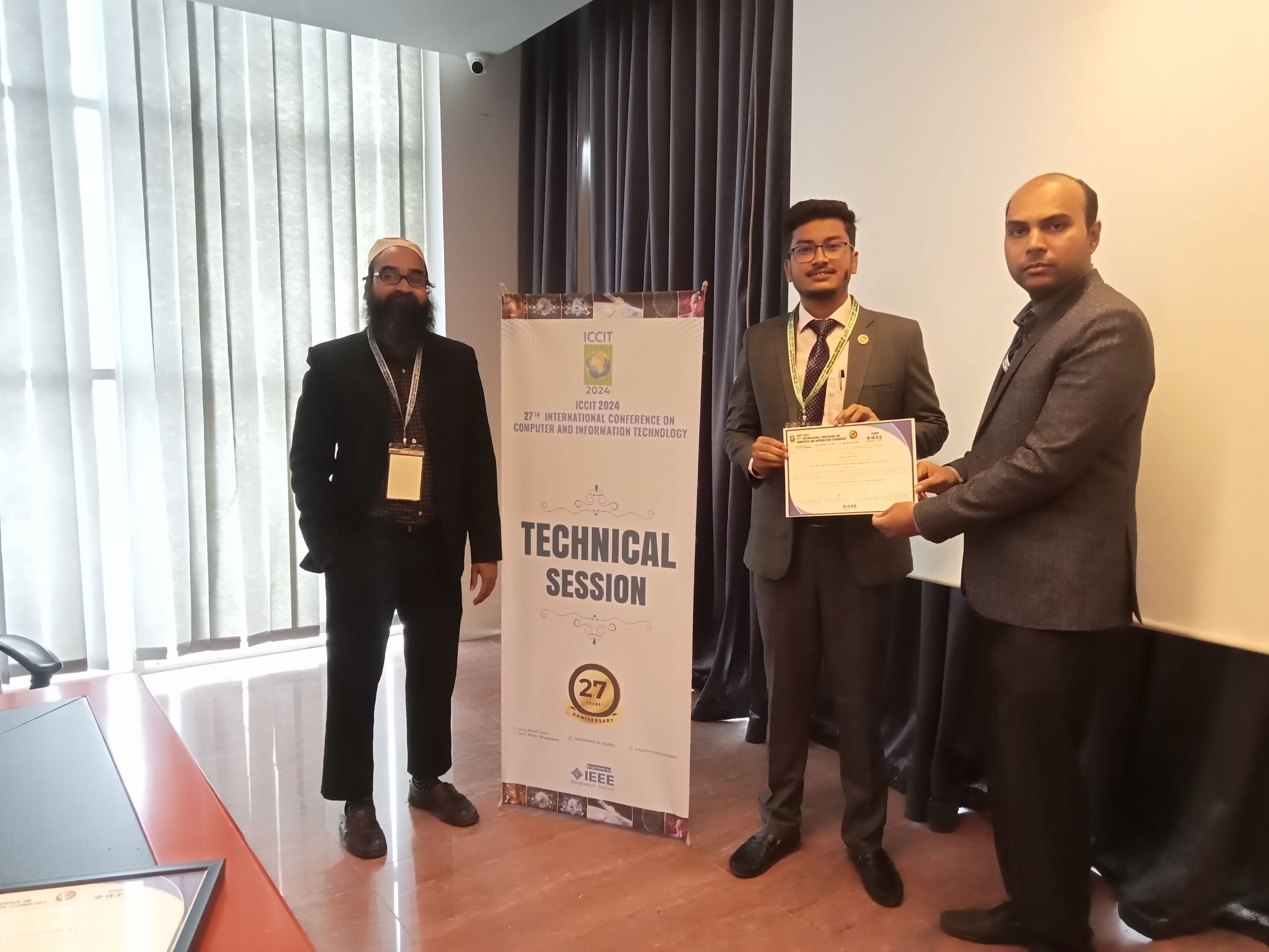
After Technical Session

After closing ceremony of the conference at Long Beach Hotel, Cox's Bazar

